


[Year:2019] [Month:May-June] [Volume:10] [Number:3] [Pages:1] [Pages No:165 - 165]
DOI: 10.5005/jp-journals-10015-1641 | Open Access | How to cite |

[Year:2019] [Month:May-June] [Volume:10] [Number:3] [Pages:4] [Pages No:166 - 169]
Keywords: Bulk-fill composite, Fracture resistance, Short fiber composite, Sonic activation
DOI: 10.5005/jp-journals-10015-1637 | Open Access | How to cite |
Abstract
Background: Class II MOD cavity in maxillary premolar creates a specific challenge for the restoration material in terms of longevity and fracture resistance due to the anatomical shape of premolars that render them susceptible to fracture and the microleakage issue of composite restoration at the gingival margin of the proximal boxes. Bulk-fill composite was introduced to provide more strength and resistance and also to provide less polymerization shrinkage and better cure depth. With the advances in dental material science and technology, several attempts have also been made to increase the advantage of bulk-fill composite: by modifying the monomers, utilizing special restoration placement instrument, and adding fiber reinforcement to its composition, which have not been compared adequately. Hence, this study was undertaken to evaluate the effect of different bulk-fill composites in class II MOD cavities on upper premolars in terms of fracture resistance. Materials and methods: A total of 30 sound upper premolars were divided into three groups of 10 each. Teeth were prepared in the form of class II MOD cavity and restored accordingly: group I restored with Filtek bulk-fill (3M), group II with Sonicfill bulk-fill (Kerr), and group III with EverX bulk-fill (GC). Afterward, samples were thermocycled at 5°C and 55°C for 500 cycles. Fracture resistance test was done using Torsee's Electronic System Universal Testing Machine. Data obtained were analyzed with one-way ANOVA and post hoc least significant difference (LSD) test to determine the difference between groups. Results: ANOVA statistical test showed no significant differences (p > 0.05) in all groups. However, resin composite EverX bulk-fill (GC) has a higher fracture resistance (882.94 ± 64.41 N) compared to other groups, Sonicfill bulk-fill (Kerr) (856.48 ± 101.35 N), and Filtek bulk-fill (3M) (812.15 ± 66.89 N). Conclusion: The use of different bulk-fill resin composites did not yield significant effects in terms of fracture resistance in the restoration of class II MOD cavity on upper premolars (p > 0.05). However, bulk-fill resin composite did offer advantages in clinical applications due to the simplified restoration process and reduced working time.

[Year:2019] [Month:May-June] [Volume:10] [Number:3] [Pages:7] [Pages No:170 - 176]
Keywords: Bone loss, Immunization, Periodontitis, Porphyromonas gingivalis, Rats
DOI: 10.5005/jp-journals-10015-1639 | Open Access | How to cite |
Abstract
Aim: Periodontitis is an inflammatory disease causing destruction of tooth-supporting structures. It is often caused by gram-negative microorganisms such as Porphyromonas gingivalis (P. gingivalis). Common treatments for periodontitis are often nonspecific and include mechanical plaque removal and surgery. This study aimed to assess the amount of bone loss and antibody titer against P. gingivalis in rats. Materials and methods: This in vitro experimental study was conducted on 66 Surrey rats free of black pigmented pathogens, which were randomly divided into six groups of 11. Groups I and II were vaccinated with formalin-killed whole-cell (FKWC) P. gingivalis with incomplete Freund's adjuvant as the vaccine carrier, and groups III and IV were vaccinated with incomplete Freund's adjuvant and PG buffer. Groups V and VI were considered as positive and negative controls, respectively. Three weeks later, they were vaccinated with a booster dose. At 28 days, groups I, III, and V were inoculated with viable P. gingivalis (ATCC 33277) four times at 48-hour intervals for induction of periodontitis. One week after booster dose administration and two weeks after oral inoculation of bacteria, serum and saliva samples were obtained for assessment of antibody titer. Ten weeks after final bacterial inoculation, the serum and saliva samples were obtained to assess antibody titer, and subgingival plaque samples were obtained from the maxillary second molar site to assess the bacterial count. The rats were then sacrificed to assess bone loss. Results: Serum and saliva antibody titers in groups I and II were significantly different from those in other groups one week after booster dose and two and 10 weeks after oral inoculation of bacteria (p < 0.001). In terms of bone loss and bacterial count in the subgingival plaque, group I was not significantly different from the negative control group and groups II, IV, and VI (p > 0.99), but had a significant difference with the positive control (group V) and group III (p < 0.001). Conclusion: This study showed successful immunization against P. gingivalis, which increased serum IgG and saliva IgA titers, limited the colonization of P. gingivalis in subgingival plaque, and restricted the alveolar bone loss.

[Year:2019] [Month:May-June] [Volume:10] [Number:3] [Pages:4] [Pages No:177 - 180]
Keywords: Double blind, LLLT, Ni-Ti closed coil spring, Randomized control clinical trial, Temporary anchorage device
DOI: 10.5005/jp-journals-10015-1633 | Open Access | How to cite |
Abstract
Aim: The aim of the present study was to assess the effect of low level laser therapy in increasing the rate of orthodontic tooth movement. Materials and methods: Twenty-four arches in 24 patients above 18 years of age requiring bilateral extractions in the same arch were randomly selected for this study. By this way, both the patient and the postgraduate student were blinded in the study. The experimental side was exposed to biostimulation using 980 nm gallium–aluminum–arsenide (GaAlAs) diode lasers, and the contralateral side was taken as control. Laser irradiation was delivered with a power output of 2 W in a continuous wave mode. The laser beam was delivered using a 1 × 4 cm diameter tip held perpendicular and in contact with the mucosa at the cervical third of canine on the buccal and palatal surfaces over an area of 4 cm2. Digital caliper measurements accurate to ±0.001 mm were recorded on study cast models on the 1st day, 28th day, 57th day, and 85th day. The distance between the contact points of the maxillary canine and second premolar was measured on study cast models three times, and the mean value was used for data computations. Results: On comparison of the rate of tooth movement between the control and laser groups, the tooth movement was greater in the laser group than in the control group, and it was statistically highly significant at all time intervals with the level of significance set at 0.05 at 95% confidence interval. Conclusion: LLLT with a specified regimen applied once in a month is effective in increasing the rate of orthodontic tooth movement.

[Year:2019] [Month:May-June] [Volume:10] [Number:3] [Pages:5] [Pages No:181 - 185]
Keywords: CT scanning, Maxillofacial, Maxillomandibular trauma, Ultrasonography
DOI: 10.5005/jp-journals-10015-1632 | Open Access | How to cite |
Abstract
Background: Head and neck trauma forms a major proportion of patients requiring maxillofacial care. Imaging is a vital part in the management of these patients. Many modalities exist that maybe utilized for the purpose of visualizing the fracture defects. Computed tomography (CT) scans have been considered the gold standard even with many disadvantages like associated radiation hazards, high cost factor, unavailability in rural healthcare setup, and time taken for the scans. Aim: Through our study, we aim to compare the efficacy of ultrasonography (USG) and compare it with CT scan in the detection of maxillomandibular fractures. Materials and methods: Over a 2-year period, 50 patients suspected with zygomatico-complex (ZMC) or mandible fractures underwent clinical examination followed by radiographic assessment using ultrasound and CT scans. The sensitivity, specificity, and time taken to evaluate the fractures were determined and compared. Results: High correlation existed between USG and CT scan in the detection of the maxillomandibular fractures. The sensitivity was 95% and the specificity was 90% with a high positive predictive value. Conclusion: Apart from a few disadvantages like difficulty in interpretation of a film without a report or clinical correlation and examiner variability, the advantages associated with its use probably outweigh the shortcomings.
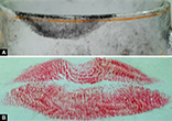
Study on Lip Prints—To Estimate the Reliability as a Personal Identification Method
[Year:2019] [Month:May-June] [Volume:10] [Number:3] [Pages:6] [Pages No:186 - 191]
Keywords: Biometrics, Fingerprints, Forensic odontology, Lip prints
DOI: 10.5005/jp-journals-10015-1629 | Open Access | How to cite |
Abstract
Aim: Lip prints are unique for any individual. The potential of lip prints as one of the biometric records to determine the identity has been well-recognized. However, studies focused on their reliability by comparing the developed latent lip prints were scarce. This study focused on the reliability of the lip prints for the personal identification by comparing the registered lip prints with the developed latent lip prints made on the porcelain cups. Materials and methods: Samples of 102 subjects (52 males and 50 females) within the age group of 18–30 years were included. Latent and superimposed lip impressions were made on a standard porcelain cup. The latent prints were developed with the fingerprint powder. Then, the lip prints with lip rouge were registered on a transparent adhesive tape. Both the developed latent lip prints and the registered lip prints were photographed with a standard ruler using the digital camera and were compared. The lip prints were classified with the scheme proposed by Tsuchihashi. The statistical analysis was done using the Pearson Chi-square test (IBM SPSS version 20) with a p value of 0.05. Results: The lip prints were unique to any individual irrespective of the gender variation. Their interpretation of comparing the digital pictures confirmed the presence of unique patterns and the possibility of the feature extraction similar to the fingerprints. Type III was the most frequent pattern observed in the study group. Conclusion: We conclude that the lip prints are highly reliable as a biometric record due to their uniqueness. The lip prints have demonstrated enough evidence that is intentionally registered and the developed latent prints were compared, which can be applied as one of the easiest and simplest methods for comparison. Yet, the authenticity of the lip print is in the preliminary level and need more systematic studies to be accepted for the legal disputes. Clinical significance: The study result can strengthen the reliability of the lip prints as an identification tool and discusses the future possibilities of lip print application.

[Year:2019] [Month:May-June] [Volume:10] [Number:3] [Pages:5] [Pages No:192 - 196]
Keywords: Arabic coffee, Caffeinated beverages, Provisional composite restorative materials, Stimulating drinks
DOI: 10.5005/jp-journals-10015-1628 | Open Access | How to cite |
Abstract
Aim: The objective of the study is to examine the effects of the consumption of caffeinated drinks common to the Saudi population regarding the mechanical properties of composite resin restorative provisional materials. Materials and methods: An in vitro approach has been utilized to analyze the flexural strength and microhardness of three different composite provisional restorative materials: Temphase™, Protemp™, and CAD Temp® monoColor, when immersed in three different caffeinated drinks: Arabic coffee, American black coffee, and cappuccino, distilled water was used as the control group for a duration of seven days (n = 10 for each test). Results were analyzed using two-way ANOVA and Tukey HSD tests. Results: All beverages significantly reduced the flexural strength of different composite provisional restorative materials investigated in the study. All beverages significantly reduced the microhardness of the Temphase™ material. Arabic coffee did not significantly affect the microhardness values of the Protemp™ material and did not have an impact on the microhardness of the CAD Temp® material. Conclusion: The surface microhardness and flexural strength of different composite resin materials were altered after emersion in different caffeinated drinks. Clinical significance: As excessive consumption of caffeinated beverages have a negative effect on the durability and longevity of the different composite resin provisional restorative materials, dentists should counsel patients with provisional restorations to reduce the detrimental effect associated with excessive consumption of these beverages.

Facial Fractures in Preschool- and School-aged Children
[Year:2019] [Month:May-June] [Volume:10] [Number:3] [Pages:5] [Pages No:197 - 201]
Keywords: Facial fractures, Maxillofacial fractures, Maxillofacial injuries
DOI: 10.5005/jp-journals-10015-1640 | Open Access | How to cite |
Abstract
Aim: The purpose of this study was to analyze the patterns of facial fractures in children and to compare them between preschool- and school-aged children. Materials and methods: This retrospective observational study included 57 children with facial fractures. The variables analyzed were the age of the patients—divided into a preschool-aged group (0–5 years) and a school-aged group (6–12 years)—gender, cause of trauma, the facial bones involved, the pattern of fracture, the modality of treatment used, the time between injury and treatment, and the postoperative complications. Results: The incidence of facial fractures in children ≤12 years was 30.2%. The patients consisted of 40 (70.2%) males and 17 (29.8%) females, and most patients belonged to the school-aged group (n = 35, 61.4%). The most common cause of injury was falls. Mandibular fractures were the most common (54.2%), mostly involving the condylar region. Forty patients (70.2%) were treated surgically and 17 patients (29.8%) were managed conservatively. The variables that were significantly different between the two groups included the cause of injury, the site of injury, and the type of treatment. Conclusion: Facial fractures occur most frequently in school-aged children with male predominance, falls are the most common cause of facial fractures in children, the incidence of mandibular fractures is high and the condyle is the most affected site, the surgical treatment is indicated in most of the older age groups, and no major complications were encountered. Clinical significance: Facial fractures in children require special considerations in their management due to many characteristic features of the facial skeleton of the growing child and the possibility of growth disturbances that may result from these injuries, the incidence of facial fractures in children increases with the beginning of school and their treatment in school-aged children tends to be surgical rather than conservative.

[Year:2019] [Month:May-June] [Volume:10] [Number:3] [Pages:5] [Pages No:202 - 206]
Keywords: Dentin hypersensitivity, Diode laser, Fluoride varnish, Iontophoresis
DOI: 10.5005/jp-journals-10015-1626 | Open Access | How to cite |
Abstract
Aim: The aim of the present study was to assess the efficacy of three different treatment modalities in the management of dentinal hypersensitivity (DH). Materials and methods: A split-mouth, randomized clinical study with a total of 30 (12 males and 18 females) patients aged between 18 years and 45 years, with 60 hypersensitive teeth, was conducted. The extent of DH was assessed using the tactile stimuli test and cold air blast. A total of 60 hypersensitive teeth from 30 patients were randomly divided into three groups: group I, hypersensitive teeth treated with iontophoresis with acidulated phosphate fluoride (APF) gel; group II, hypersensitive teeth treated with a diode laser (DL); and group III, hypersensitive teeth treated with fluoride varnish. All the three groups were assessed for DH using visual analogue scale (VAS) at baseline and on days 15 and 30 after the initial treatment. An analysis of variance (ANOVA) repeated measure analysis was done and p < 0.05 was considered as statistically significant. Results: Out of the 30 enrolled patients, 18 (60%) were females and 12 (40%) were males. Most of them (43.3%) were aged between 36 and 45 years. A majority of the included teeth were anterior [32(53.3%)]. The mean VAS scores among three groups for tactile stimulus response showed a maximum reduction (1.90 ± 1.20) of DH in group II with a DL after 30 days. Similar reductions were recorded in the mean VAS scores among three groups for cold air blast stimulus response, with maximum reduction (2.02 ± 0.10) of DH in group II after 30 days. A statistically significant difference was observed among all the three groups. Conclusion: The present study concluded that a 980 nm wavelength DL demonstrated significantly greater reduction of DH followed by iontophoresis with APF and fluoride varnish, respectively.

[Year:2019] [Month:May-June] [Volume:10] [Number:3] [Pages:7] [Pages No:207 - 213]
Keywords: 17% EDTA, 0.2% high molecular chitosan solution, 2.5% NaOCl, iRace, ProTaper Universal rotary file
DOI: 10.5005/jp-journals-10015-1638 | Open Access | How to cite |
Abstract
Aim: To compare the effect 2.5% NaOCl, 17% ethylenediaminetetraacetic acid (EDTA), and 0.2% high molecular chitosan solution during root canal preparation using ProTaper Universal and iRace rotary file on corrosion of NiTi instrument and quantity of apically extruded nickel ions. Materials and methods: Twenty-four extracted human premolars were divided into 4 groups. Group I was the sample with root canal preparation using ProTaper Universal rotary file with 2.5% NaOCl and 17% EDTA, group II was the sample using ProTaper Universal rotary file with 2.5% NaOCl and 0.2% high molecular chitosan solution, group III was the sample using iRace rotary file with 2.5% NaOCl and 17% EDTA, and group IV was the sample using iRace rotary file with 2.5% NaOCl and 0.2% high molecular chitosan solution. After root canal preparation, the quantity of apically extruded nickel ions was evaluated using atomic absorption spectrophotometry (AAS). Instruments were evaluated for corrosion and surface defect using a scanning electron microscope. Results: A combination of 2.5% NaOCl and 0.2% high molecular chitosan solution caused significantly lower corrosion and surface defect in ProTaper Universal and iRace rotary files and also produced fewer extrusions of nickel ions compared to a combination of 2.5% NaOCl and 17% EDTA. The mean value of corrosion and surface defect scores on iRace was lower and also resulted in less extrusion of nickel ions compared to ProTaper Universal rotary. Conclusion: 0.2% high molecular chitosan solution can be developed as a chelating material, an alternative to EDTA.

[Year:2019] [Month:May-June] [Volume:10] [Number:3] [Pages:5] [Pages No:214 - 218]
Keywords: Double blind, Initial archwire, LLLT, Randomized control trial, VAS
DOI: 10.5005/jp-journals-10015-1634 | Open Access | How to cite |
Abstract
Aim: To compare and evaluate the relative analgesic efficacy of low-level single-dose laser and placebo irradiation after the placement of the first arch wire (0.016 inch Ni–Ti wire) at 6, 12, 24, 36, 48, and 72 hours using a visual analog scale (VAS). Materials and methods: Fifteen maxillary arches of 15 patients were selected for the study. All the brackets and molar tubes were bonded and 0.016-inch nickel–titanium wire was inserted and ligated to the bracket using 0.009 inch stainless steel wire. After performing the orthodontic procedures, a low-level laser therapy (LLLT) of wavelength of 980 nm an output of 2 W with an energy density of 40 J/cm2 side, and an application dose of 10 J/cm2 were applied on the buccal and palatal sides of each tooth for 20 seconds, respectively. The laser beam was placed for the control group on the contralateral side for the same extent of time but in the switched off mode. Patients were asked to assess the pain at home at 6, 12, 24, 36, 48, and 72 hours after irradiation using a VAS. Results: In both the groups, pain increased during the first 12 hours and gradually decreased over the next couple of days. The peak of pain was maximum at 12 hours post irradiation in both the groups. There was a statistically significant (p < 0.005) reduction in pain in the experimental group than the control group at all-time intervals. Conclusion: LLLT significantly reduces orthodontic tooth pain due to the initial archwire activation.

[Year:2019] [Month:May-June] [Volume:10] [Number:3] [Pages:8] [Pages No:219 - 226]
Keywords: Constant taper, Endodontic concepts, Variable taper, Working length, Working width
DOI: 10.5005/jp-journals-10015-1635 | Open Access | How to cite |
Abstract
Aim: The present study aims to assess the knowledge of some endodontic concepts, working width (WW) vs working length (WL) and variable taper vs constant taper among endodontic practitioners. Materials and methods: A descriptive study design was employed to assess the knowledge and understanding of endodontics practitioners. The questionnaire was distributed among 400 participants, which was composed of different items and was measured using different rating scales. Results: Descriptive statistics were presented in tables based on the frequency and percentages of responses from participants. Out of 400 participants, 363 responses were obtained. The results showed that the radiographic method and the electronic method were both useful for determining the WL during root canal treatment. Moreover, a majority of the participants preferred to use WL with 0.5 mm shorter than apex, while a few of them preferred 1 mm shorter than the apex. The significant factors that help in determining the WW and length included canal taper, canal morphology, and canal curvature. Conclusion: The study results have concluded that both the radiographic method and the electronic method are useful in determining the WL. Canal geometry and morphology were among the factors that influence the WL detection method.

[Year:2019] [Month:May-June] [Volume:10] [Number:3] [Pages:8] [Pages No:227 - 234]
Keywords: Contemplation, Modified-Fagerstrom rating questionnaire scale (m-FTQ), Nicotine dependence, Pre-contemplation, Smokers
DOI: 10.5005/jp-journals-10015-1627 | Open Access | How to cite |
Abstract
Nicotine dependence is a challenging health problem presently among smokers as it involves a compulsive use of a substance in spite of its known harmful effects. Aim: The primary aim is to assess the nicotine dependence in a group of smokers by using seven-question m-FTQ (modified Fagerstrom questionnaire) and to assess the nicotine dependence with age, duration in years, and the number of packets smoked per day. Materials and methods: Only those who use smoking forms of tobacco in the age groups between 16 years and 79 years were randomly selected by visiting Vinayaka Mission's Sankarachariyar Dental College, Vinayaka Missions Research Foundation. The total sample size is 200 patients. The age groups were divided into < 20, 21–30, 31–40, 41–50, 51–60, 61–70, and 71–80. Informed consents were obtained from the participants of the study. The questionnaire was carried at chairside in the outpatient Department of Oral Medicine, Diagnosis and Radiology, Vinayaka Missions Sankarachariyar Dental College, Vinayaka Missions Research Foundation, Salem. Results: Beedi smokers were at a risk of developing nicotine dependence when compared to cigarette and churut smokers. Conclusion: Churut smokers were found to have high mean nicotine dependence Fagerstrom score than beedi and cigarette smokers. In this study, moderate nicotine dependence Fagerstrom score was found to be predominant among the smoking population of Salem. Clinical significance: This study helps to assess nicotine dependence among smokers in Salem and the necessity to provide counseling for the cessation of tobacco and establishment of tobacco cessation centers.

[Year:2019] [Month:May-June] [Volume:10] [Number:3] [Pages:6] [Pages No:235 - 240]
Keywords: Adolescent, Bruxism, Dental caries, Habits, Malocclusion, Quality of life
DOI: 10.5005/jp-journals-10015-1631 | Open Access | How to cite |
Abstract
Aim: The aim of the present study was to assess the impact of bruxism on oral health related quality of life (OHRQoL) among 12 to 15-year-old school-going children in Mangaluru. Materials and methods: A case-control study was conducted among 108 12 to 15 year-old school-going children in Mangaluru. Cases were identified from children having poor OHRQoL and controls as children having better OHRQoL by selecting median scores using Child Perception Questionnaire (CPQ11–14). Bruxism was identified using the criteria of American Association of Sleep Medicine (AASM) questionnaire. Information regarding dental caries, dental wear, and malocclusion were also collected. Results: Bruxism was significantly associated with OHRQoL subscales such as emotional and social well-being scores, respectively (p = 0.01 and p = 0.02). Malocclusion also showed statistically significant association with OHRQoL subscale of emotional well-being score. However, there were no significantly significant associations between bruxism and overall mean OHRQoL scores (p > 0.05). Conclusion: Bruxism had statistically significant association with OHRQoL subscales, and was shown to have significant impacts on social and emotional wellbeing of participants. Results indicate that bruxism might affect the psychosocial functioning of children. Clinical significance: Bruxism is a common clinical condition encountered by clinicians. The results of the present study indicate that it can have a definite and deeper impact on children's emotional health and also on the way they socialize. It can thus be one of the critical predictors of psychosocial functioning of the children.

Implications of Plasma Lymphoblastic Cells in Lymphoreticular Disorders: An Overview
[Year:2019] [Month:May-June] [Volume:10] [Number:3] [Pages:5] [Pages No:241 - 245]
Keywords: Hematopoietic stem cells, Immunoglobulins, Lymphoreticular system, Lymphoblasts, Plasmablasts
DOI: 10.5005/jp-journals-10015-1636 | Open Access | How to cite |
Abstract
Aim: The aim of this review was to emphasize the diverse morphologic features of plasma lymphoblastic cells in lymphoreticular disorders to arrive at a precise diagnosis. Background: The lymphoreticular system comprises of a group of cells with a common lineage and primary function of immunoregulation. Specific immunity is achieved by the combined effects of macrophages and lymphocytes, and, therefore, it is the lymphoreticular system. These cells are scattered in different parts of the body and share some functional characteristics. At both functional and anatomical levels, lymphoreticular tissue can be categorized into primary and secondary lymphoid organs that predominantly produce lymphocytes and plasma cells. Review results: The plasma lymphoblastic lesions/malignancies comprise of characteristic cells like buttock cells, cells with irregular nuclei, cells with cleaved nuclear outlines, etc. Identification of such cells amidst sheets of malignant lymphoblastic cells is challenging. However, sound knowledge about the morphology of these cells and their immunohistochemical panel of markers may provide a clue for diagnosis. Conclusion: The predominant cell types noted in plasma lymphoblastic lesions histopathologically are immature lymphocytes and plasma cells in their varied cell activity suggest the biologic behavior of the lesion. Clinical significance: Understanding and identifying the normal and pathological cellular and nuclear morphology of the lymphoreticular cells can aid in the definitive diagnosis of the plasma lymphoblastic disorders and predict its biological nature.

Qualitative Research and its Role in Public Health Dentistry
[Year:2019] [Month:May-June] [Volume:10] [Number:3] [Pages:5] [Pages No:246 - 250]
Keywords: Evidence-based dentistry, Mixed methods, Qualitative research, Research designs
DOI: 10.5005/jp-journals-10015-1630 | Open Access | How to cite |
Abstract
Aim: The purpose of this review was to comprehensively explore various aspects of qualitative research, its methods, applications, challenges, and recommendations pertaining to dentistry. Background: Qualitative research is a form of social inquiry concerned primarily with how people see and understand their social world. It has hypothesized the field of social sciences and proved to be helpful in health sciences as well. It has a descent from anthropology and philosophy to sociology. Evidence shows that this method was first used by anthropologists and sociologists back in the early decades of 20th century, as a method of inquiry. The qualitative research cycle consists of three interlinked methods: the design cycle, ethnographic cycle, and analytical cycle. In-depth interviews, focus group discussions, observation, and documentary analysis are methods to collect data. Most commonly practiced methods are the first two approaches. An approach of investigation involving both quantitative and qualitative data analysis is called as mixed methods research. Review results: Literature search was done with the help of Endnote software, followed by systematically arranging the retrieved articles in a synchronized manner. Few studies were found in the field of public health dentistry, which employed a qualitative approach to probe into a few sensitive issues. Conclusion: Although the future scope of qualitative research in this field is immense, it continues to be underutilized. Qualitative research complements quantitative research work as it explores complex phenomena and areas in which quantitative research alone may not be amenable to.


© Jaypee Brothers Medical Publishers (P) LTD.

